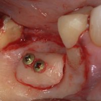
This patient presents with a buccal bone deformity that resulted from a previous extraction. The bone may have been compromised from a prior infection from periodontal disease and or from the removal of the tooth. The patient desires to have this tooth replaced by a dental implant.
A narrow diameter dental implant could be used in this situation. However the resulting crown (restoration) will be compromised by being over contoured. It will have to taper from the biting surface down to approximately 3mm instead of 5-6mm that would be necessary for a normal emergence profile. Not only does this create food traps but it is also difficult for the patients to cleanse the areas where the restoration is over contoured. These areas frequently get neglected which can lead to halitosis, gingival inflammation and possibly periimplantitis. Bio-mechanically, excess torque is placed on the implant due the discrepancy of the width of the biting surface of the tooth relative to the neck of the tooth that must taper dramatically to meet the narrow implant interface. The discrepancy creates a cantilever.
Another option is to augment the deformed ridge via bone grafting to allow a normal diameter implant for a molar restoration which is in the 5-6mm range. A block bone graft is the preferred method for this procedure, and is performed by a periodontist.
The lateral Ramus, shown in the picture gallery above, is a good donor site for the block graft. 3D cone-beam tomography is necessary to map the location of the mandibular nerve as well as visualize the donor and recipient sites of the block graft.
Once the block graft is removed it is shaped and contoured to the approximate dimensions of the bony defect. The block bone graft is then secured to the recipient site by lag screws. These screws are typically removed months later when the implant is placed. While there can be some gaps or voids, these are filled by particulate bone which is used to complete the contours.
In this case, platelet rich plasma was also used to enhance the healing. The particulate bone, mineralized freeze dried bone, was saturated with platelet rich plasma prior to use. The collagen membranes used to cover the grafts of both the donor and recipient sites are also saturated in platelet rich plasma prior to placement.
The gingival flaps were advanced to cover the newly grafted areas. This resulted in the molar being covered by part of the gingival which can easily be corrected at a future appointment, if the tissue doesn’t shrink back on its own.
Pain medications, anti-inflammatory agents, antibiotics and aseptic mouth rinses are prescribed following this procedure. There is moderate discomfort that is controlled well with medications. The primary post operative sequelae is post surgical swelling.
The implant is typically placed after 4-6 months after the grafting procedure.


No comments yet.