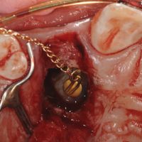
Abnormal eruption of a maxillary canine can result in an impacted tooth, where the tooth can no longer erupt into its proper position in the arch. The tooth gets physically stuck in the bone where other tooth roots block its passage through the bone. This case demonstrates a maxillary impacted canine, tooth #6 which has erupted horizontally, rather than vertically. Subsequently, the crown of tooth #6 ran into the root of the central incisor, tooth #8, which is now blocking it.
Periodontal surgery can be performed to expose the crown of the tooth. From there, a bracket is bonded to the tooth. The tooth can then be orthodontically maneuvered back into its correct position in the dental arch. This surgery is a form of crown lengthening.
One of the challenges with this procedure is determining orientation of the impacted tooth relative to the tooth root that is blocking its path. This orientation is necessary so that the Periodontist knows whether to approach the surgical exposure from the lip (buccal) or roof of the mouth (palatal) aspect. 3D cone beam tomography is employed to determine this orientation. As you can see from the 3D renderings, it was determined that the crown of the impacted #6 was palatal to the root of #8 which is blocking its eruption path. Thus the tooth must be accessed from the palatal aspect as it would not be possible or could severely damage the root of #8 by trying to orthodontically move it in the buccal direction.
A palatal gum flap is reflected. Initially bone was covering the crown of impacted tooth #6. Bone is gently removed in the area where the crown of the impacted tooth is suspected to be. The bone proves to be very thin in that area and is easily penetrated. The follicle surrounding the crown is quickly exposed. Its like a balloon sack around the crown of any erupting tooth. The follicle is removed and the area is debrided. This exposes the crown of the impacted tooth.
An orthodontic bracket is bonded to the newly exposed crown. A laser is used to create a hole in the gum flap to allow the chain connected to the bracket to exit out of the tissue. The exposed crown will also follow this path as the tension from the chain guides the tooth back into its proper position into the dental arch. The chain is initially ligated to the arch wire. The patient is then referred back to their orthodontist to have the chain attached to the orthodontic appliance so that tension can be applied.
The process of erupting the tooth happens over weeks to months. Generally the deeper and more horizontal the impaction, the longer it will take to erupt back into the arch.


How long will it take approximately to get the canine into it’s original (if the tooth was in the same place as the pictures above).
I’m eighteen and I have this problem! I wish I could avoid having braces so instead I’m trying to prepare myself!
Many thanks, Kate.
Depending on how impacted it is about 4-12 months.
my daughter is 13 years with an impacted canine, but the overlying bone has resorbed up to the distal of the lateral incisor. the apex of the canine is almost formed. there is a plan to do bone augmentation on the resorbed area before guiding the canines eruption. What is the prognosis for this procedure? thank you very much
Without doing a clinical evaluation and seeing the radiographs it is difficult to say. In general I would move the canine first before doing a bone graft. If the bone resorbed in the first place you would have to question the wisdom of trying a bone graft for the same situation without moving the canine first.
my 10 yrs old will have this done both upper canines are impacted. honestly, how painful is this? what is the recovery time?
It depends on if the canines will be exposed toward the lip or toward the roof of the mouth. But in general its about 7-10 days of healing and Kids heal much quicker than adults. Typical pain meds for a procedure like this are ibuprofen (Motrin) and Hydrocodone (Vicodin). Most would get by with just ibuprofen.
what ada codes would this be under
Unfortunately, I am not sure, but I will try to find out for you