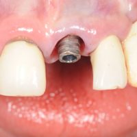
Guided dental implant surgery affords such precision and predictability that we were able to use the patient’s own extracted tooth as a temporary restoration. This is not a reason in and of itself to use guided implant surgery its just perk of using the technique. Immediate extraction and placement of a dental implant in the maxillary anterior area is very difficult. The maxilla is thin and it is very easy for the operator to perforate the bone during an immediate extraction case because gingival flaps are typically not reflected. Also the slope of the bone from the extraction socket tends to force the drill bit toward the apex of the extraction socket which will direct the drill outside of the bony housing if the drill goes in that orientation. Prior to guided surgery the Periodontist would orient the drill toward the palatal aspect, but again there is not a lot of margin of error before perforating the palatal plate of bone. Palatal orientation will also necessitate the use of an angulated final abutment to compensate for the palatal orientation of the implant. Guided surgery eliminates the problems above. Using specialized software and a 3D tomography a dental implant image can be transposed into the radiographic image. Once the optimal placement is achieved, a surgical guide can be fabricated to duplicate the exact placement in the human body. Because the guide stabilizes and orients the drilling sequence the resulting bony slope from the extraction socket is no longer a factor in deflecting the bone drills.
In this case, it was possible to predict that the resulting implant placement would allow for the possibility of using the patients own tooth to be used as a temporary restoration. The drilling sequence and implant placement went as planned using the surgical guide. A temporary abutment was placed and the height was reduced to allow for the temporary restoration. Most of the root portion was resected from the crown of the tooth. From there, the crown was hollowed out, per the pictures above. This left a shell of the tooth. The shell portion is filled with a special acrylic then placed over the abutment. Flash acrylic is carefully removed. The tooth is set slightly lower and slightly buccal to its original placement so that there is no initial tooth contact from the opposing tooth. Even with guided surgery this is still not a very predictable procedure, there can be issues with the original tooth or trauma from the extraction that my preclude its use.
When the extracted tooth can be used as a temporary, it provides for a very bio-compatible restoration that should preserve the original gum architecture. Connective tissue attachment can happen to the remaining root portion of the tooth. The gingival architecture is also supported by the original tooth. Overall there should be minimal dimensional changes. Cosmetically the patient is left unchanged.
I would not advocate using the original tooth crown as a permanent restoration, though modifications could be made to this technique to improve its long term use. The shell of the original tooth is fragile and still susceptible to tooth decay. It is not difficult to replicate the esthetics’s and contours of the original tooth using zirconia abutments and all ceramic restorations and dentists have been doing this very successfully for quite some time now.
This patient will use this restoration for the next 2-4 months till the implant has successfully integrated. Once that happens this restoration will be removed and a permanent restoration will be fabricated.


No comments yet.